Blood Vessels in the Knee: A Labeled Diagram of Arteries and Veins
Blood vessels in the knee: a labeled diagram of the popliteal artery, genicular arteries and knee veins, plus knee and calf signs that need urgent care.
The main blood vessels in the knee are the popliteal artery and the popliteal vein, which run together through the shallow hollow at the back of the knee (the popliteal fossa). The popliteal artery continues from the femoral artery, the main artery of the thigh, after it passes through an opening in a thigh muscle. It sends five genicular branches around the knee joint, then divides below the knee into the arteries of the lower leg. [1] The popliteal vein forms from the deep veins of the calf, receives the small saphenous vein and carries blood up into the thigh, where it is called the femoral vein. [1] [2] Another vein, the great saphenous vein, runs under the skin on the inner side of the knee. [3] [4] Arteries take blood away from the heart; veins bring it back. [5]
Safety first: call 911 or your local emergency number if a leg turns cold, pale, numb or weak, or becomes hard to move, which can be a sign of a blocked artery. [6] Call 911 if leg swelling comes with shortness of breath or chest pain. If you have been told you have a clot in your leg, go to the emergency room or call 911 for chest pain, coughing up blood, difficulty breathing or fainting. [7] [8] Call a healthcare provider right away for swelling, pain, tenderness, warmth or red or discolored skin in a leg, or for pain, swelling, numbness, tingling or bluish color in the calf below a sore knee. [9] [10] This page explains normal anatomy. It cannot tell you whether you have a clot, a cyst or an artery problem.
This page describes typical adult anatomy in general terms. Branching differs from person to person, most often in the branches of the popliteal artery, and some people have a doubled popliteal vein. [1] [2] It is not a guide to an individual’s blood vessels, symptoms or test results.
The major blood vessels around the knee
The table lists the named vessels in the diagram: arteries from the thigh down, then veins from the calf up.
| Vessel | Kind | Where it runs | What it does |
|---|---|---|---|
| Popliteal artery | Main artery | Through the back of the knee, deeper than the popliteal vein. [1] [2] | Continues from the femoral artery and is the main blood supply to the back of the knee and the lower leg. [1] [2] |
| Genicular arteries: superior medial, superior lateral, middle, inferior medial, inferior lateral | Branches of the popliteal artery | Above, behind and below the knee joint; the middle one goes into the joint. [1] | Supply the joint capsule and ligaments, and link up into a network around the knee and kneecap. [1] [11] |
| Descending genicular artery | Branch of the femoral artery | Starts in the thigh, above the knee, from the inner wall of the femoral artery. [3] [11] | Its articular branch joins the network around the kneecap; its saphenous branch supplies the skin over the front and inner side of the knee. [11] [3] |
| Anterior tibial, posterior tibial and fibular (peroneal) arteries | Arteries of the lower leg | Begin just below the knee, where the popliteal artery divides into the anterior tibial artery and a shared trunk for the other two. [1] | Carry blood on into the lower leg; small recurrent branches of the anterior tibial artery loop back up to the knee network. [11] [4] |
| Popliteal vein | Deep vein | Through the back of the knee, beside the artery and closer to the skin. [1] [2] | Forms from the anterior tibial, posterior tibial and fibular veins, carries blood from the lower leg toward the heart and becomes the femoral vein in the thigh. [1] [2] |
| Small saphenous vein | Superficial vein | Up the back of the calf, coming out between the two heads of the calf muscle (gastrocnemius). [4] | Drains the back of the lower leg into the popliteal vein. [2] |
| Great saphenous vein | Superficial vein | Under the skin on the inner side of the knee, toward the back. [4] [3] | Returns blood from the surface of the leg and drains into the femoral vein through an opening high in the thigh (the saphenous opening). [3] |
Deep veins run among the muscles beside the arteries, and superficial veins lie under the skin. Short perforating veins pass through the layer of tissue between them and connect the two. [4] The tibial nerve runs through the same space, closer to the skin than the vessels. It is a nerve, not a blood vessel. [1]
Labeled diagram: arteries and veins at the back of the knee
Both panels show the back of a right knee, as if you were standing behind the person: the inner side is on the left and the outer side on the right. Panel 1 follows the arteries down; panel 2 follows the veins back up. Labels, arrows and line styles carry the meaning, so you do not need to tell colors apart to read it. [1] [4]
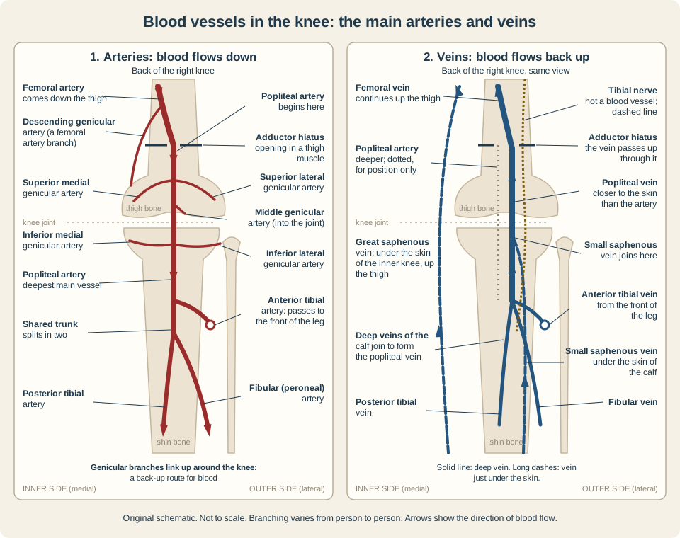
Full text equivalent
- Panel 1, arteries: the femoral artery comes down the thigh and passes through an opening in the adductor magnus muscle (the adductor hiatus). Below the opening it is called the popliteal artery. [1]
- Above the opening, the femoral artery gives off the descending genicular artery, which sends branches to the knee. [11] [4]
- Behind the knee, the popliteal artery gives off five genicular arteries: superior medial, superior lateral, middle, inferior medial and inferior lateral. The middle genicular artery supplies the ligaments and lining inside the joint. [1]
- These branches link up with one another around the knee, giving blood an alternative route from the femoral artery to the popliteal artery. [1] [4]
- Below the knee, the popliteal artery divides into the anterior tibial artery, which heads to the front of the leg, and a shared trunk that splits into the posterior tibial and fibular (peroneal) arteries. [1] [2]
- Panel 2, veins: the anterior tibial, posterior tibial and fibular veins from the calf join to form the popliteal vein. [1]
- The popliteal vein runs up the back of the knee beside the popliteal artery and closer to the skin. The artery appears in this panel as a thin dotted line, for position only. [1]
- The small saphenous vein comes up under the skin of the calf and joins the popliteal vein behind the knee. [4] [2]
- The popliteal vein passes up through the adductor hiatus and becomes the femoral vein. [1] [2]
- The great saphenous vein runs under the skin of the inner knee and up the thigh to the femoral vein. [3] [4]
- The tibial nerve, drawn as a short-dashed line, runs through the same space. It is not a blood vessel. [1]
How blood moves through the knee
Arteries take blood away from the heart, so blood in the popliteal artery flows down toward the lower leg. Veins bring blood back, so blood in the popliteal vein flows up toward the heart. [5] [2]
- Back-up routes. The network of genicular arteries gives blood an alternative path from the femoral artery to the popliteal artery. [1]
- Calf muscle pump. When the calf muscles contract, pressure in the lower-leg veins rises, and blood moves up into the popliteal vein and back toward the heart. [2]
- Deep and superficial veins. Perforating veins connect the veins under the skin with the deep veins. [4]
Normal anatomy versus unexplained pain or swelling
Knowing where the vessels run does not tell you why a knee hurts or swells. Knee pain can have many causes, including problems in the joint itself such as arthritis, a torn ligament or a meniscus tear. [10] Several problems at the back of the knee can look alike from the outside, which is why clinicians examine and test rather than going by location alone. [12]
- Baker cyst. A fluid-filled swelling behind the knee that often comes with arthritis or a meniscus tear. Often it causes no symptoms. If it breaks open, it can cause pain, swelling and bruising of the knee and calf, which a blood clot can also cause. MedlinePlus says it is important to know which one it is, because a clot needs medical attention right away. [10] [13]
- Blood clot in the popliteal vein. The popliteal vein is one of the most common places for a deep vein thrombosis (DVT). [2] Symptoms can include swelling, pain or tenderness, cramping, aching or warmth, and red or discolored skin. [9] A clot can break loose and travel to the lungs (pulmonary embolism). [14] Common signs of that include shortness of breath, pain with deep breathing, rapid breathing and a faster heart rate. [15]
- Popliteal artery aneurysm. A bulge in the wall of the popliteal artery. It is the most common true aneurysm among the peripheral arteries, often shows up as a pulsing lump at or above the back of the knee, and is confirmed with an ultrasound scan. [1]
- Blocked artery. A leg that turns cold, pale, numb or weak, or is hard to move, can mean an artery is blocked. MedlinePlus says to go to the emergency room or call 911. [6]
What a diagram cannot tell you
A clot in a vein sometimes causes no obvious signs. [14] Clinicians diagnose DVT from symptoms, medical history, a physical exam, and imaging or blood tests; an ultrasound of the leg looks for clots in the deep veins. [9] [8] No home check can rule a clot in or out, so bring new or unexplained knee or calf pain or swelling to a healthcare professional rather than matching it to a drawing.
For more on how blood moves through the body, browse Heart and circulation or use the circulatory system worksheets to practice naming arteries and veins. To practice checking what a food or supplement label does and does not say, try the food label reading worksheet.
Common questions
What is the main artery behind the knee?
The popliteal artery. It continues from the femoral artery after the adductor hiatus and is the main blood supply to the knee region and the lower leg. [1] [2]
Is the popliteal vein a deep vein?
Yes. It is a deep vein of the leg, although it also receives a superficial vein, the small saphenous vein. [2]
Why does the knee have so many small arteries?
The genicular arteries supply the joint capsule and ligaments. Because they link up with one another, they also give blood an alternative route around the knee. [1]
Is a swelling behind the knee always a Baker cyst?
No. A blood clot, other cysts or lumps, and a popliteal artery aneurysm can look similar, so a clinician needs to tell them apart. [12] Contact your provider if a swelling behind the knee becomes large or painful. [13] If leg swelling comes with shortness of breath or chest pain, call 911 or your local emergency number right away. [7]
Can a blood clot form in the vein behind the knee?
Yes. The popliteal vein is one of the most common places for a DVT. [2] Call your healthcare provider right away if you think you may have symptoms of DVT. [9]
Safety first: call 911 or your local emergency number for a leg that turns cold, pale, numb or weak, or for leg swelling with shortness of breath or chest pain. [6] [7] Call a healthcare provider right away for new swelling, pain or tenderness in one leg or calf. Do not use this diagram to rule out a clot. [9] [7]
Sources and limitations
- StatPearls: Anatomy, Bony Pelvis and Lower Limb: Popliteal Region (Hyland S, Sinkler MA, Varacallo MA). Updated July 25, 2023. Anatomy reference for health professionals; describes typical anatomy and common variants, not any one person.
- StatPearls: Anatomy, Bony Pelvis and Lower Limb: Popliteal Vein (Sawyer E, Sinkler MA, Tadi P). Updated August 8, 2023. Anatomy reference for health professionals; its treatment remarks are written for clinicians.
- StatPearls: Anatomy, Bony Pelvis and Lower Limb: Saphenous Nerve, Artery, and Vein (Mathew K, Varacallo MA). Updated July 24, 2023. Anatomy reference; the course of superficial veins varies between people.
- StatPearls: Anatomy, Bony Pelvis and Lower Limb, Knee (Gupton M, Imonugo O, Black AC, Launico MV, Terreberry RR). Updated November 5, 2023. General anatomy reference; vessel positions are simplified and vary between people.
- NHLBI: How Blood Flows through the Heart. Updated June 25, 2025. General anatomy education.
- MedlinePlus Medical Encyclopedia: Arterial embolism. Review date April 28, 2026. General adult information; symptoms can begin quickly or slowly.
- MedlinePlus Medical Encyclopedia: Foot, leg, and ankle swelling. Review date May 19, 2025. General adult information; lists common causes and is not a diagnostic tool.
- MedlinePlus Medical Encyclopedia: Deep vein thrombosis. Review date April 1, 2025. General adult information; not a diagnostic tool.
- NHLBI: Deep Vein Thrombosis (DVT). Updated September 19, 2022. Symptoms vary and are not a home rule-out checklist.
- MedlinePlus Medical Encyclopedia: Knee pain. Review date November 7, 2024. General adult information; lists common causes and is not a diagnostic tool.
- StatPearls: Anatomy, Bony Pelvis and Lower Limb: Arteries (Arias DG, Marappa-Ganeshan R). Updated December 9, 2023. Describes the fibular artery as a branch of the posterior tibial artery; other chapters name a shared trunk for the same branching.
- StatPearls: Baker’s Cyst (Leib AD, Roshan A, Foris LA, Varacallo MA). Updated August 4, 2023. Written for clinicians; the exam signs it names are not home tests.
- MedlinePlus Medical Encyclopedia: Baker cyst. Review date June 27, 2026. General adult information; not a diagnostic tool.
- NHLBI: What Is Venous Thromboembolism?. Updated September 19, 2022. General clinical education.
- NHLBI: Pulmonary Embolism (PE). Updated September 19, 2022. General clinical education.
These statements have not been evaluated by the Food and Drug Administration. This product is not intended to diagnose, treat, cure or prevent any disease.
Not Medical AdviceThe information on this site is for educational purposes only and is not medical advice. Talk with a qualified healthcare practitioner before making changes to your health routine.