Types of Vein: Superficial, Deep and Pulmonary Veins
Compare superficial, deep and pulmonary veins with a labeled circulation diagram, and learn why vein appearance cannot establish a diagnosis.
Veins carry blood toward the heart. Superficial veins lie near the skin, deep veins lie deeper among tissues such as muscles, and pulmonary veins carry oxygen-rich blood from the lungs to the heart. These labels describe different things: superficial and deep refer to location; pulmonary refers to the lung circulation. They are not three mutually exclusive categories covering every vein. [1] [2]
Symptoms come before the diagram. Contact a healthcare professional right away for a swollen, tender, painful leg or suspected blood-clot symptoms. Shortness of breath or pain when breathing also requires immediate medical attention; do not use vein appearance or this diagram to decide whether care is needed. Symptoms vary, and some clots do not cause symptoms. [3] [4]
Compare the vein types
This comparison describes typical circulation after birth, with limb veins used to explain superficial and deep locations. It is not a guide to fetal circulation, congenital differences, or an individual’s anatomy. Sources are general anatomy references, not a study establishing someone’s health status. [1] [2]
| Vein type | Where it is found | What it does | Important distinction |
|---|---|---|---|
| Superficial | Near the surface, beneath the skin | Returns blood from outer tissues; connects with the deep system | Being visible does not establish a diagnosis. [2] |
| Deep | Deeper in the limbs, including among muscles | Carries blood back toward the heart | Cannot be assessed simply by looking at the skin. [2] |
| Perforating | Between superficial and deep vein systems | Connects the systems; in the normal leg, directs flow toward deep veins | A connecting route, not a separate lung circuit. [2] |
| Pulmonary | Between the lungs and the left atrium of the heart | Returns oxygen-rich blood from the lungs | A vein is defined by flow toward the heart, not by low oxygen. [1] |
Labeled circulation diagram
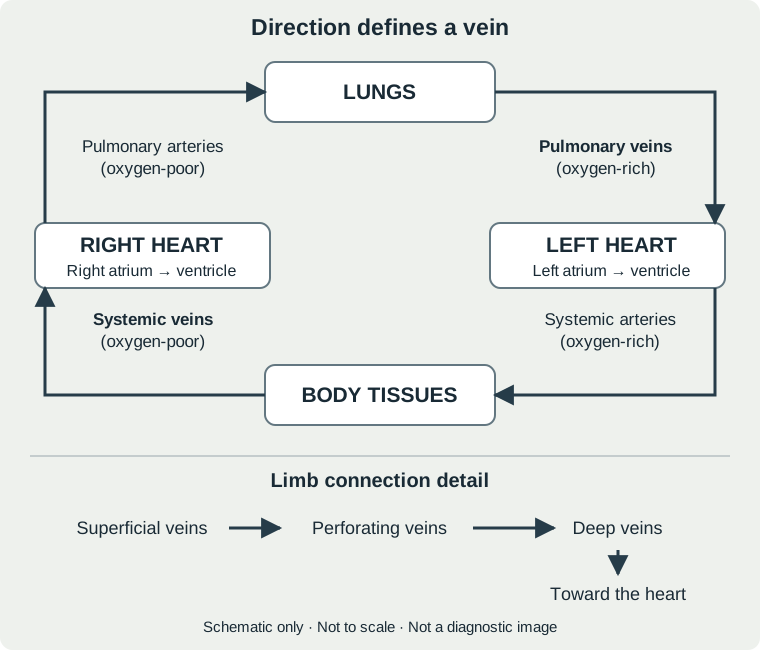
Follow the arrows to see how the body and lung circuits connect. This original schematic shows direction, not exact vessel shapes, size, branching, or position. The separate lower panel illustrates normal limb-vein connections. [1] [2]
Full text equivalent
- Blood returns from body tissues through systemic veins, including the venae cavae, to the right atrium and then the right ventricle.
- The right ventricle pumps blood through the pulmonary arteries to the lungs, where it receives oxygen.
- Pulmonary veins return oxygen-rich blood to the left atrium and then the left ventricle.
- The left ventricle pumps blood through the aorta and systemic arteries toward body tissues; blood then returns through the veins.
- In the separate limb detail, superficial veins connect through perforating veins with deep veins. Blood continues toward the heart. This detail is not a depiction of every drainage route.
Flow sequence: [1]. Limb connections: [2].
What appearance cannot tell you
A visible vessel is not a home test for a clot, healthy circulation, or oxygen level. Vein concerns may require a history, examination, and tests such as ultrasound. The absence of an obvious surface change does not rule out a problem; NHLBI notes that venous thromboembolism can occur without symptoms until complications develop. [2] [3]
Common questions
Are pulmonary veins different from pulmonary arteries?
Yes. In typical circulation after birth, pulmonary arteries carry oxygen-poor blood from the right heart to the lungs. Pulmonary veins carry oxygen-rich blood back to the left heart. Both names describe direction relative to the heart. [1]
Are perforating veins the same as deep veins?
No. Perforating veins connect the superficial and deep vein systems. The lower diagram panel separates that connecting route from the deeper veins receiving the blood. [2]
Can this diagram tell me why a vein looks different?
No. It illustrates normal flow, not a person’s symptoms or test results. A new concern should be assessed by a healthcare professional, not matched to a drawing. [2]
If you have suspected clot symptoms, seek medical attention rather than using this resource to rule out a problem. Shortness of breath or pain when breathing needs immediate attention. [3] [4]
Sources and limitations
- NHLBI: How Blood Flows through the Heart. Updated June 25, 2025. General anatomy education; not an individual diagnostic assessment.
- Cleveland Clinic: Veins. Updated June 19, 2022. General anatomy and clinical overview; not a population-specific comparative study.
- NHLBI: Venous Thromboembolism Symptoms. Updated September 19, 2022. General symptom guidance; symptoms vary and are not a home rule-out checklist.
- NHLBI: Pulmonary Embolism. Updated September 19, 2022. General clinical education.
These statements have not been evaluated by the Food and Drug Administration. This product is not intended to diagnose, treat, cure or prevent any disease.
Not Medical AdviceThe information on this site is for educational purposes only and is not medical advice. Talk with a qualified healthcare practitioner before making changes to your health routine.